Просмотр новости
Найдите то, что Вас интересует
 Спутники
 РН
РН SpaceX
 Роскосмос
 NASA
NASA
СпутникиРНSpaceXРоскосмосNASABackground Fluoride administration plays a critical role in the management and prophylaxis of dental caries. Research indicates that laser irradiation can enhance the uptake of fluoride into dental enamel, an effect that is particularly pronounced when used as an adjunct to topical fluoride treatments. The present study aimed to evaluate the impact of CO2 laser irradiation on fluoride uptake in the enamel of permanent teeth. Methods Six human upper premolars, extracted for orthodontic reasons, were selected for this study. The roots were separated from the crowns, and each crown was then sectioned mesiodistally to create mesial and distal halves. These halves were assigned to either an experimental or a control group (n = 6 per group). All specimens received an application of 5% sodium fluoride (NaF) varnish. Subsequently, the samples in the experimental group were subjected to irradiation with a 1 W CO2 laser for a duration of 15 seconds. To assess the effects of the treatments, fluoride uptake was measured and the topographic characteristics of the enamel surface were examined using scanning electron microscopy (SEM) and energy-dispersive X-ray spectroscopy (EDX). The data obtained from the split-tooth design (paired observations from the same tooth) were analyzed using non-parametric methods. The Wilcoxon Signed-Rank Test was employed to compare the chemical element concentrations (F, Ca, P, C) between the experimental (mesial) and control (distal) halves. Effect sizes (r) and 95% confidence intervals (CI) were calculated for each paired comparison. Statistical significance was set at p < 0.05. All analyses were performed using SPSS software version 26 (IBM, USA). Results Scanning electron microscopy (SEM) structural analysis revealed that the enamel surfaces in the fluoride alone group maintained a typical etched pattern with visible micropores. In contrast, the experimental group subjected to both sodium fluoride varnish and CO2 laser irradiation demonstrated distinct surface melting, fusing of enamel crystals, and a smoother recrystallized morphology. Energy-dispersive X-ray spectrometry analysis showed a significant increase in the percentage of fluoride with the application of the laser, The results of the mean ranks showed that the fluoride and CO2 laser group recorded significantly higher ranks in the fluoride element (8.75 vs. 4.25), reflecting a higher concentration and confirmed statistical significance. Statistically, the Wilcoxon Signed-Rank test revealed a significant increase in fluoride concentrations in the laser-treated group compared to the control group (P value = 0.026), which indicates the effectiveness of using CO2 lasers in enhancing fluoride absorption within the tooth structure. Conclusion The combination of CO2 laser irradiation and sodium fluoride varnish resulted in observable enamel surface morphological modifications and increased surface elemental concentrations of fluoride, calcium, and phosphorus in vitro. However, these surface alterations do not constitute direct evidence of enhanced acid resistance, fluorapatite formation, or clinical caries prevention. Further long-term clinical studies are mandatory to validate any potential therapeutic benefit.
Research Article
Revised
[version 3; peer review: 1 approved, 2 approved with reservations, 1 not approved]
Sham Mohamad Farouq Alkhateeb1, Chaza Nader Kouchaji2, Abdulrhman Ahmad Alkhaled
https://orcid.org/0000-0002-5510-4339
2, Omar Hamadah1,3Sham Mohamad Farouq Alkhateeb1, Chaza Nader Kouchaji2, Abdulrhman Ahmad Alkhaled
https://orcid.org/0000-0002-5510-4339
2, Omar Hamadah1,31 Higher Institute of Laser Research and Applications, Damascus University, Damascus, Syria
2 Department of Pediatric Dentistry, Damascus University Faculty of Dentistry, Damascus, Syria
3 Oral Medicine Department, Damascus University Faculty of Dentistry, Damascus, Syria
Sham Mohamad Farouq Alkhateeb
Roles: Data Curation, Formal Analysis, Investigation, Methodology, Software, Validation, Visualization, Writing – Original Draft Preparation, Writing – Review & Editing
Chaza Nader Kouchaji
Roles: Conceptualization, Formal Analysis, Investigation, Methodology, Software, Supervision, Validation, Visualization, Writing – Original Draft Preparation, Writing – Review & Editing
Abdulrhman Ahmad Alkhaled
Roles: Conceptualization, Data Curation, Formal Analysis, Investigation, Methodology, Resources, Software, Validation, Visualization, Writing – Original Draft Preparation, Writing – Review & Editing
Omar Hamadah
Roles: Data Curation, Formal Analysis, Investigation, Methodology, Software, Validation, Writing – Original Draft Preparation, Writing – Review & Editing
OPEN PEER REVIEW
REVIEWER STATUS
Corresponding author: Abdulrhman Ahmad Alkhaled Competing interests: No competing interests were disclosed.
Grant information: The author(s) declared that no grants were involved in supporting this work.
Copyright: © 2026 Alkhateeb SMF et al. This is an open access article distributed under the terms of the Creative Commons Attribution License, which permits unrestricted use, distribution, and reproduction in any medium, provided the original work is properly cited. How to cite: Alkhateeb SMF, Kouchaji CN, Alkhaled AA and Hamadah O. Effect of Combined CO₂ Laser Irradiation and Sodium Fluoride Varnish on Enamel Surface Morphology and Surface Elemental Composition: An In Vitro Split-Tooth Study [version 3; peer review: 1 approved, 2 approved with reservations, 1 not approved]. F1000Research 2026, 15:588 (https://doi.org/10.12688/f1000research.171971.3) First published: 19 Apr 2026, 15:588 (https://doi.org/10.12688/f1000research.171971.1) Latest published: 23 Jul 2026, 15:588 (https://doi.org/10.12688/f1000research.171971.3)
Changes between V2 and V3:
This revised version addresses all concerns raised by the peer reviewers. The major changes are as follows:
1. Statistical analysis: The Mann-Whitney U test was replaced with the Wilcoxon Signed-Rank test, which is the appropriate paired statistical method for our split-tooth design where each tooth served as its own control.
2. Specimen allocation: Inconsistencies regarding randomization were resolved. The Methods now clearly state that mesial halves were consistently assigned to the experimental group (fluoride + laser) and distal halves to the control group (fluoride only).
3. Methodological details: We added a detailed description of varnish removal using acetone-soaked cotton pellets followed by ultrasonic cleaning in distilled water. We also explicitly described examiner blinding, where all specimens were assigned coded identification numbers to mask group assignment from SEM and EDX operators.
4. Interpretational caution: The Abstract, Discussion, and Conclusion were thoroughly revised to avoid over-interpretation. We now explicitly state that SEM and EDX analyses provide topographic and compositional data only, and do not constitute evidence of enhanced acid resistance, fluorapatite formation, or clinical caries prevention. A dedicated limitation paragraph was added to the Discussion.
5. Data presentation: Effect sizes (r) and 95% confidence intervals were added to the Wilcoxon test results. A footnote was added to Table 6 clarifying the interpretation of Z-scores and effect size calculations.
6. Duplication removal: A repeated paragraph in the Discussion was removed.
These revisions have substantially improved the scientific rigor, methodological transparency, and interpretational accuracy of the manuscript.
See the authors' detailed response to the review by Sanpreet Singh Sachdev
Dental caries deserves attention because it has become a chronic disease affecting children worldwide.1,2 Dental caries is a multifactorial disease caused by the simultaneous interaction of various factors— sugar and dental biofilms —in the oral cavity. The introduction of fluoride into dental practice over seventy years ago is widely regarded as the principal reason for the global decline in dental caries. Despite this public health success, the sole confirmed adverse effect of fluoride use in dentistry is dental fluorosis, a condition resulting from excessive fluoride ingestion during tooth development. Recent epidemiological trends indicate a concurrent rise in the prevalence of dental fluorosis and a decline in caries rates.3
The most common varnish used in dentistry to prevent tooth caries is sodium fluoride (NaF).4 Topical application of NaF on teeth mitigates enamel demineralization while enhancing the remineralization process. The primary antimicrobial mechanism of fluoride is not direct bacterial destruction, but rather an indirect action through the inhibition of bacterial acid production, which results in a higher pH at the tooth surface.5,6
The combined use of laser irradiation and topical fluoride represents a novel approach to caries prevention by increasing the enamel’s affinity for and incorporation of fluoride. Zancope et al. reported that CO2 lasers cause morphological changes on the surface of tooth enamel, such as fusion and dissolution.7 Studies evaluating the effect of carbon dioxide laser on the structure of tooth enamel and dentin have shown high absorption by dental tissue.8 Specifically, the 10,600 nm CO2 laser features an absorption coefficient in hydroxyapatite that allows specific depth of penetration and flow to produce the desired thermal effects on caries inhibition.9
Although topical fluoride applications, such as varnishes, remain the gold standard in caries prevention, their clinical efficacy is often limited by their susceptibility to rapid dissolution in oral fluids, necessitating frequent reapplications. However, repeated high-dose applications raise concerns regarding potential systemic toxicity and fluorosis.3 To overcome these limitations and enhance the longevity of fluoride retention, modern preventive dentistry has focused on synergistic technologies. The CO2 laser (10,600 nm) has emerged as a promising modality; when irradiated at specific sub-ablative parameters, it induces microstructural and thermal alterations in the enamel surface. This photothermal effect temporarily increases enamel permeability and promotes the stable conversion of hydroxyapatite into a highly acid-resistant fluorapatite phase.10 Therefore, the integration of laser therapy is proposed not to replace fluoride, but to optimize its chemical binding and depth of penetration, thereby potentially offering prolonged cariostatic protection with lower effective fluoride doses.
This study aimed to evaluate and compare the efficacy of CO2 laser irradiation versus a CO2 application in enhancing fluoride uptake by the surfaces of permanent teeth following treatment with NaF varnish.
The six human upper premolars were obtained from the Department of Oral and Maxillofacial Surgery in College of Dentistry, University of Damascus after routine extractions for orthodontic reasons and not specifically for this study. Teeth were stored in distilled water at 4 °C until use.
Teeth were examined to ensure they were free of any caries, abrasions or fractures. They were divided into two sections, a mesial section and a distal section, so that each treatment was applied to a section of the tooth. The root was cut from the Cementoenamel junction and the crown were stored in distilled water to prevent dehydration ( Figure 1).
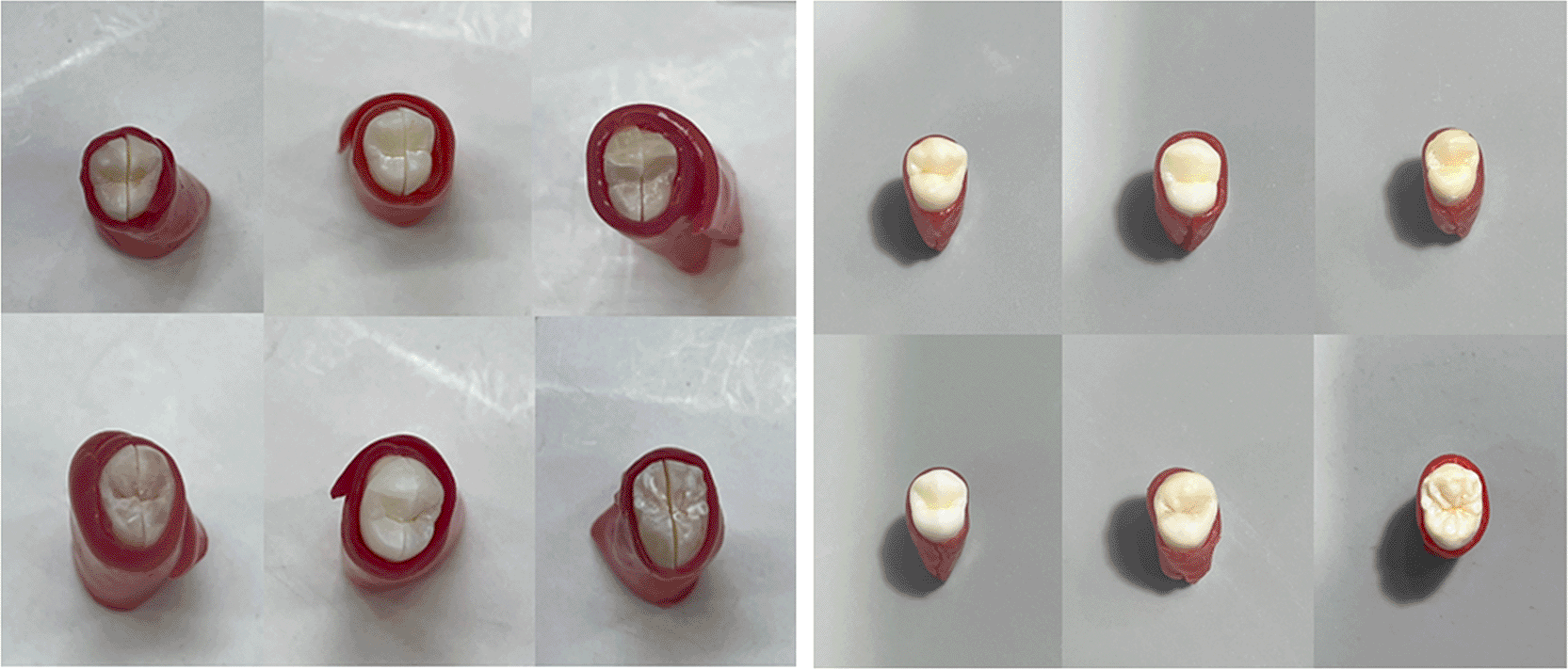
Mesial section was used as the experimental specimen and the distal as the control.
All specimens were first dried and treated with 5% NaF varnish (VOCO®, Germany). A distinction was then made between the groups: the experimental specimens were laser irradiated, while the control specimens were not. Sodium fluoride varnish (5% NaF varnish (VOCO, Germany, Product Code: [190/10]) was applied to the mesial section of the tooth with a carbon dioxide laser with a wavelength of 10600 nm (power 1 W – pulse duration 15 ms) while the control sample was in the distal section and sodium fluoride varnish alone was applied ( Figure 2).
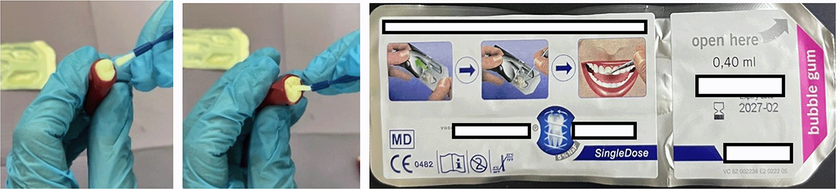
The varnish was applied using a small brush according to the manufacturer’s instructions by applying a thin layer over the entire occlusal surface of the tooth.
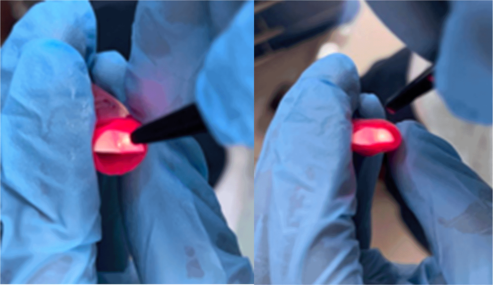
The sample in the mesial section was exposed to CO2 laser radiation (10600 nm wavelength, 1 W power, pulsed mode, 15 ms pulse duration) with a wavelength of 10600 nm and a power of 1 W for 15 seconds with a pulsed mode and a pulse duration of 15 ms11 at a distance of 1 cm. Irradiation was performed in a continuous scanning motion that allowed the entire surface to be irradiated and we re-preserved it with distilled water ( Figure 3).
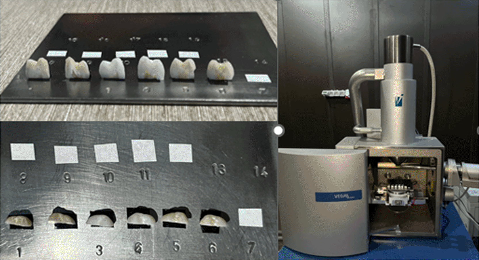
The test of the effect of the laser on the varnish was evaluated using scanning electron microscopy (SEM) (T-Scan company VEGA II XMU, Czech Republic). Prior to SEM and EDX analyses, the remaining fluoride varnish was thoroughly and systematically removed from the enamel surfaces. Each specimen was gently wiped for 30 seconds using fresh cotton pellets saturated with pure acetone to dissolve the organic matrix of the varnish, followed by an immersion in an ultrasonic bath of distilled water for 5 minutes to eliminate any remaining chemical residues. Finally, the surfaces were thoroughly rinsed with a continuous stream of distilled water for 60 seconds and dried using a oil-free air syringe. The amount of fluoride, calcium, phosphorus, and carbon remaining on the specimens was tested using energy-dispersive X-ray spectroscopy (EDX) (Oxford Instruments, UK) attached to the microscope located in the Atomic Energy Commission ( Figure 4). To ensure reproducibility and eliminate bias, examiner blinding was implemented during the microscopic evaluation. All specimens were assigned coded identification numbers by a primary investigator, completely masking the group assignment (experimental vs. control) from the SEM and EDX operators. Furthermore, the EDX acquisition protocol was standardized utilizing an accelerating voltage of 20 kV with a fixed working distance across all analyzed specimens. The samples were analyzed by SEM to observe the topographic properties of the enamel surface after different treatments, and images of the central region were taken using two different detectors: Secondary Electrons (SE) and Backscattered Electrons (BSE). EDX analysis provided non-destructive quantitative data on the chemical composition and elemental distribution over the surface area.12 The response variables evaluated were changes in the concentrations of fluoride, carbonate, calcium, and phosphorus that constitute the hydroxyapatite structure.
This in vitro study utilized extracted human teeth collected from patients who underwent orthodontic treatment. This study was from 24/8/2025 to 27/9/2025. The study protocol was reviewed and approved by the Institutional Review Board (IRB) of College of Dentistry, University of Damascus On August 21, 2025 under reference number (DN-210-125-3-9-1/21/8/2025). All participants (or their guardians) provided written informed consent for the use of their extracted teeth for research purposes, in accordance with the principles of the Declaration of Helsinki. Patient identifiers were removed to ensure confidentiality.
The results were evaluated according to the surface shape in electron microscope images and according to the concentration of fluoride, calcium, phosphorus and carbon.
In this study, SEM images showed that CO2 laser irradiation fuses the surface, creating a smooth, recrystallized side. Fusion between enamel surface crystals was also observed after carbon dioxide laser treatment ( Figures 5 and 6).
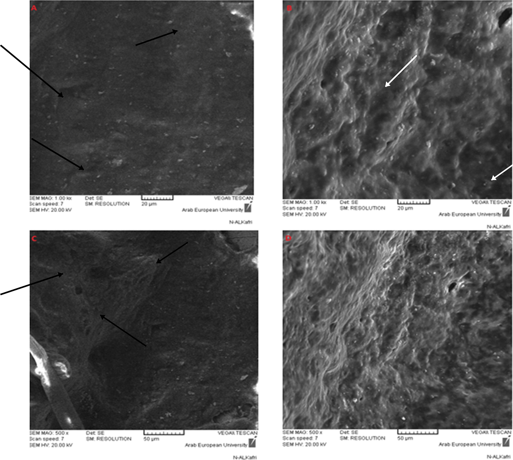
(A) Mesial section (fluoride + laser) using SE detector (×1000): Black arrows indicate zones of superficial enamel melting and crystal fusion. (B) Distal section (fluoride only) using SE detector (×1000): White arrows point to typical patent micropores. (C) Mesial section (fluoride + laser) at ×500 magnification: Black arrows highlight the smooth, recrystallized surface topography. (D) Distal section (fluoride only) at ×500 magnification showing unaltered surface morphology.
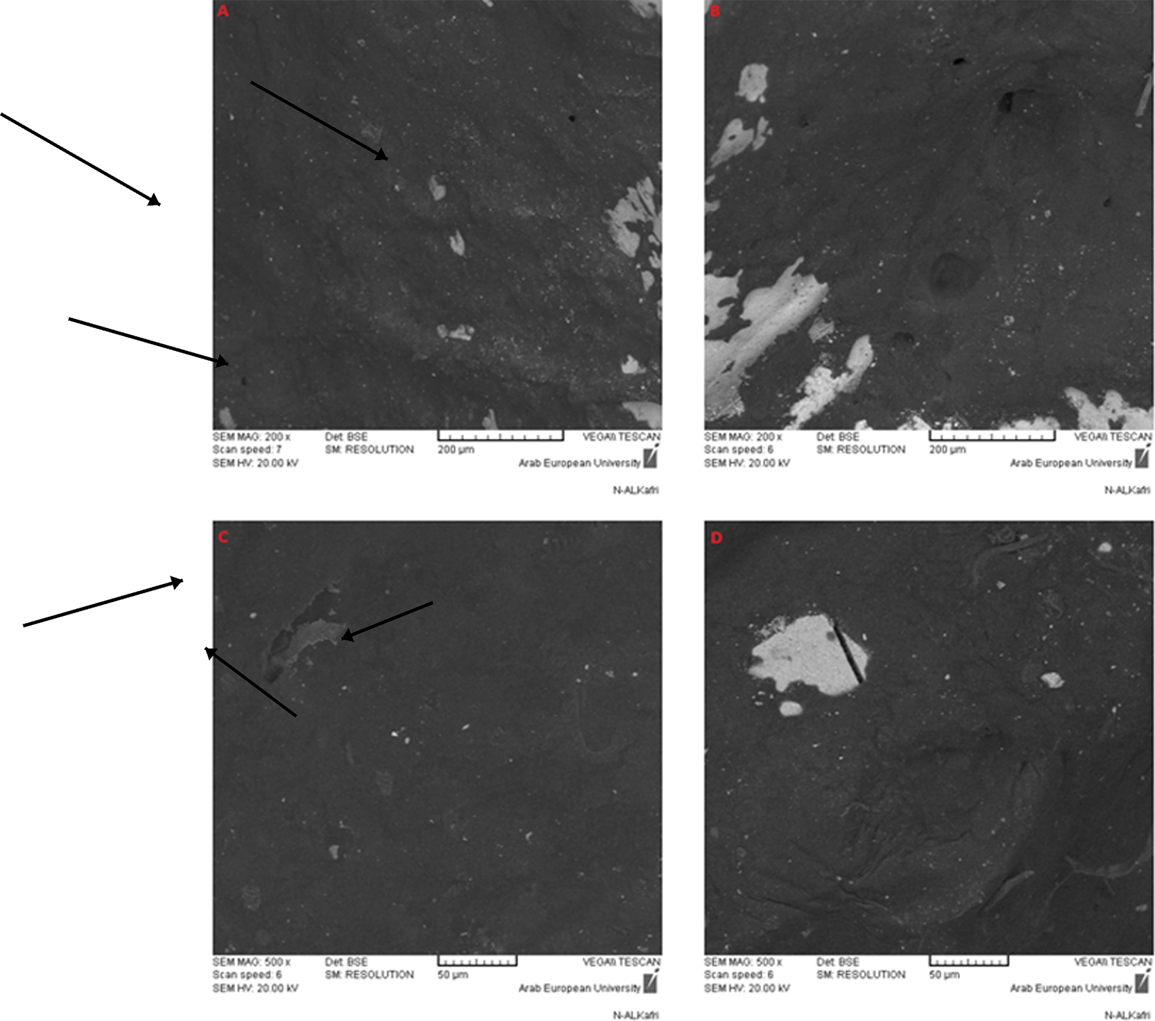
(A) Mesial section (fluoride + laser) at ×200 : Black arrows indicate the boundary of thermal modification and fused mineral deposits. (B) Lateral section (fluoride only) at ×200 showing baseline surface characteristics. (C) Medial section (fluoride + laser) at ×500 : Black arrows mark the homogeneous recrystallized hydroxyapatite lattice. (D) Lateral section (fluoride only) at ×500 displaying micro-roughness without melting features.
Comparing the apparent differences in the mean of the measured items between the two groups ( Table 1) and the descriptive statistics for fluoride, calcium, phosphorus, and carbon ratios in both study groups ( Tables 2–5).
Test values represent Wilcoxon signed-rank Z-scores. Negative Z-values indicate the direction of the difference between paired observations (laser-treated mesial halves vs. control distal halves). Effect sizes (r) were calculated as r = |Z|/√n, where n = 6 paired observations. All comparisons were based on paired data from the same tooth (split-tooth design).
The Wilcoxon Signed-Rank test revealed a statistically significant increase in fluoride, calcium, and phosphorus weight percentages in the laser-treated group compared to the control group (p < 0.05). Conversely, carbon demonstrated a significant reduction following treatment. Detailed paired differences, effect sizes (r), and 95% confidence intervals are summarized in (Table 6).
This study evaluated the efficacy of a CO2 laser for enhancing fluoride uptake in dental enamel. According to existing literature, the combined application of a CO2 laser with fluoride varnishes or gels can potentially quadruple fluoride absorption, which may significantly improve caries prevention and reduce the required fluoride dosage or frequency of clinical applications.13 While our current laboratory findings support this positive synergistic trend by demonstrating a statistically significant increase in surface fluoride concentrations (p < 0.001) via EDX analysis using the Wilcoxon Signed-Rank test, these results must be interpreted within specific boundaries.
Due to the small sample size utilized in this investigation (n = 6 permanent teeth sectioned to create 12 experimental halves), this work should be appropriately characterized as a pilot study. In advanced microstructural and surface examinations using SEM and EDX, small sample sizes are frequently employed because the focus is on highly precise, localized topographic and elemental changes rather than biological variance. Furthermore, our study utilized a split-tooth design—where each tooth served as its own internal control (mesial half treated with laser + fluoride, distal half with fluoride alone)—which effectively eliminated inter-individual biological variability and enhanced the statistical power of the comparison. Nevertheless, we acknowledge the small sample size as a limitation of this laboratory model, and future large-scale quantitative trials are required to fully validate the clinical extent of this laser-induced enhancement.
In order to obtain a protective effect, a laser was used to raise the temperature of the tooth enamel to the melting point of its inorganic components or to the decomposition point of its organic components. On the other hand, non-thermal laser radiation was used after tooth fluoridation to achieve a slow fluoride release effect. This type of reaction is called laser-activated fluoride treatment.11
Carbon dioxide laser irradiation of tooth enamel, within certain parameters, results in a specific temperature increase above the irreversible pulpitis threshold of 5.5 °C,14 which changes the chemical composition of the tooth enamel surface and the morphological structure15 and the increase may lead to permanent damage to the tooth pulp.
Excessive temperature elevation during laser application poses a risk of injury to the dental pulp. A study addressing this risk compared the thermal effects of two laser systems: a CO2 laser (at 1 and 2 W) and a diode laser (at 5 and 7 W). The results indicated that the mean temperature rise remained below the 5.5 °C critical threshold for pulp damage. Based on this evidence, the authors determined that both lasers could be used safely without compromising pulp health.16
The absorption depth of port and ivory is 12 μm, compared to the absorption depth of water of 15 μm.17
The absorption effects of tooth enamel and dentin at these depths depend on how the laser energy affects them and the laser energy depends on laser parameters such as power, mode of operation (continuous or pulsed wave mode), flow (energy density) and dose.
The continuous wave mode resulted in carbonization, dissolution and cracking of the enamel and the development of the pulse mode allowed variations by frequency and pulse duration in milliseconds (ms) and microseconds (μs). This improved the thermal control of the laser in tissue. The dose depends on the radiation method: spot radiation or sweeping motion, the number of repeated radiations.9
Several hypotheses have been advanced to explain the mechanisms by which combined laser and fluoride treatment enhances resistance to enamel demineralization. A principal theory posits that laser irradiation creates micro-spaces within the enamel structure, which subsequently serve to retain fluoride ions more effectively.18 These structural alterations are also believed to contribute directly to reducing enamel solubility.19 The observed morphological changes on the enamel surface following the sequential application of topical fluoride and CO2 laser irradiation provide a rationale for the superior protective effect compared to fluoride treatment alone.20 This synergistic effect is further attributed to the laser-induced conversion of hydroxyapatite to the more acid-resistant fluorapatite phase, a process accompanied by the melting and recrystallization of hydroxyapatite crystals.21
The results of previous studies showed that treating the surface of tooth enamel using a carbon dioxide laser reduces demineralization of tooth enamel compared to the control group. Studies have shown that wavelengths from 9 to 11 micrometers of CO2 lasers are effectively absorbed by dental hydroxyapatite, causing loss of carbonate mineral, which in turn reduces acid reactivity.22,23
The integration of lasers in preventive dentistry reflects a modern paradigm shift within the field, moving away from a traditional focus on restorative treatment and toward a philosophy of disease prevention. This approach is consistent with the recommendations of major professional bodies and is part of a wider scientific consensus advocating for less invasive caries management strategies,24 Which aims to preserve tissues to the maximum extent, and in this new approach, prevention has acquired an important role that it has not played before in the past.11
All experiments combining CO2 and fluoride laser radiation have shown better results in preventing tooth caries when compared to a single treatment. Therefore, this ‘combination therapy’ may be clinically effective, and at the same time, it may only involve moderate daily doses of fluoride and low levels of laser radiation energy, which is consistent with our current study.
A critical limitation of this study is that SEM and EDX analyses provide topographic and compositional data only. While we observed increased surface fluoride and calcium concentrations, along with surface melting and recrystallization patterns, these findings cannot be directly interpreted as evidence of fluorapatite formation, reduced enamel solubility, or enhanced acid resistance. The measured elemental changes reflect surface modifications rather than functional resistance to demineralization. Future investigations should incorporate direct acid resistance assays (e.g., pH cycling models, microhardness testing, or polarized light microscopy) to confirm whether these surface alterations translate into clinically meaningful protection against dental caries.
Within the limitations of this in vitro study, the combined application of CO2 laser and sodium fluoride varnish resulted in observable surface morphological changes (surface melting and recrystallization) and increased surface elemental concentrations of fluoride, calcium, and phosphorus compared to varnish alone. However, these topographic and compositional alterations do not constitute direct evidence of enhanced acid resistance, reduced enamel solubility, or clinical caries prevention. Long-term clinical trials incorporating direct acid resistance tests (e.g., pH cycling or microhardness testing) are mandatory to validate any potential therapeutic benefit.
Although these results are very promising, more studies should be developed in order to test these radiation conditions under different challenges, and simulate other clinical variables and therefore, future clinical studies are recommended to confirm these results.
The dataset includes raw EDX values for fluoride, calcium, phosphorus, and carbon, as well as the data used for statistical analyses and tables are It is available to the public at this link.
Figshare: Comparison between the protective effect of applying sodium fluoride varnish alone and applying CO2 laser with it in permanent teeth. https://doi.org/10.6084/m9.figshare.31333387.v225
The dataset includes:
• Research Data
• EDx images
Data are available under the terms of the CC BY 4.0
Extended data associated with this study, including EDX spectra, and detailed statistical output tables, are available in the same repository as the underlying data under a CC BY 4.0 license.
The author(s) declared that no grants were involved in supporting this work.
Published: 23 Jul 2026, 15:588
Published: 05 Jul 2026, 15:588
Published: 19 Apr 2026, 15:588
© 2026 Alkhateeb SMF et al. This is an open access article distributed under the terms of the Creative Commons Attribution License, which permits unrestricted use, distribution, and reproduction in any medium, provided the original work is properly cited.
Current Reviewer Status: ?
Key to Reviewer Statuses VIEW HIDE
ApprovedThe paper is scientifically sound in its current form and only minor, if any, improvements are suggested
Approved with reservations A number of small changes, sometimes more significant revisions are required to address specific details and improve the papers academic merit.
Not approvedFundamental flaws in the paper seriously undermine the findings and conclusions
Version 3
VERSION 3
PUBLISHED 23 Jul 2026
Revised
Reviewer Report 10 Aug 2026
Sanpreet Singh Sachdev, Oral Pathology and MIicrobiology, Bharati Vidyapeeth (Deemed to be University) Dental College and Hospital Navi Mumbai, Navi Mumbai, Maharashtra, India
Not Approved
VIEWS 0
Competing Interests: No competing interests were disclosed.
Reviewer Expertise: Oral Pathology, Dental Materials, Dental Innovations, Dental Anatomy, Dentistry
CloseReviewer Report 07 Aug 2026
Mays Sameer Tareq, University of Technology, Baghdad, Iraq
Approved with Reservations
VIEWS 0
Is the work clearly and accurately presented and does it cite the current literature?
Yes
Is the study design appropriate and is the work technically sound?
Yes
Are sufficient details of methods and analysis provided to allow replication by others?
Partly
If applicable, is the statistical analysis and its interpretation appropriate?
Partly
Are all the source data underlying the results available to ensure full reproducibility?
Yes
Are the conclusions drawn adequately supported by the results?
Partly
Competing Interests: No competing interests were disclosed.
Reviewer Expertise: laser-tissue interaction. laser ablation, laser medical applications, nanotechnology. and lasers in dentistry
CloseReviewer Report 03 Aug 2026
Vikas Singh, Teerthanker Mahaveer University, Moradabad, Uttar Pradesh, India
Approved with Reservations
VIEWS 0
Competing Interests: No competing interests were disclosed.
Reviewer Expertise: Public Health Dentistry, Preventive Dentistry, Oral Epidemiology, Clinical Research Methodology, Biostatistics, Evidence-Based Dentistry, Dental Materials
CloseVersion 2
VERSION 2
PUBLISHED 05 Jul 2026
Revised
Reviewer Report 16 Jul 2026
Vikas Singh, Teerthanker Mahaveer University, Moradabad, Uttar Pradesh, India
Not Approved
VIEWS 0
Is the work clearly and accurately presented and does it cite the current literature?
Partly
Is the study design appropriate and is the work technically sound?
No
Are sufficient details of methods and analysis provided to allow replication by others?
Partly
If applicable, is the statistical analysis and its interpretation appropriate?
No
Are all the source data underlying the results available to ensure full reproducibility?
Partly
Are the conclusions drawn adequately supported by the results?
No
Competing Interests: No competing interests were disclosed.
Reviewer Expertise: Public Health Dentistry, Preventive Dentistry, Oral Epidemiology, Clinical Research Methodology, Biostatistics, Evidence-Based Dentistry, Dental Materials
CloseReviewer Report 10 Jul 2026
Sanpreet Singh Sachdev, Oral Pathology and MIicrobiology, Bharati Vidyapeeth (Deemed to be University) Dental College and Hospital Navi Mumbai, Navi Mumbai, Maharashtra, India
Not Approved
VIEWS 0
Competing Interests: No competing interests were disclosed.
Reviewer Expertise: Oral Pathology, Dental Materials, Dental Innovations, Dental Anatomy, Dentistry
CloseVersion 1
VERSION 1
PUBLISHED 19 Apr 2026
Reviewer Report 04 Jun 2026
Sanpreet Singh Sachdev, Oral Pathology and MIicrobiology, Bharati Vidyapeeth (Deemed to be University) Dental College and Hospital Navi Mumbai, Navi Mumbai, Maharashtra, India
Not Approved
VIEWS 0
Is the work clearly and accurately presented and does it cite the current literature?
No
Is the study design appropriate and is the work technically sound?
No
Are sufficient details of methods and analysis provided to allow replication by others?
No
If applicable, is the statistical analysis and its interpretation appropriate?
I cannot comment. A qualified statistician is required.
Are all the source data underlying the results available to ensure full reproducibility?
No source data required
Are the conclusions drawn adequately supported by the results?
No
Competing Interests: No competing interests were disclosed.
Reviewer Expertise: Oral Pathology, Dental Materials, Dental Innovations, Dental Anatomy, Dentistry
CloseReviewer Report 19 May 2026
Alhan Ahmed Qasim, University of Baghdad, Baghdad, Baghdad Governorate, Iraq
Approved
VIEWS 0
Is the work clearly and accurately presented and does it cite the current literature?
Partly
Is the study design appropriate and is the work technically sound?
Yes
Are sufficient details of methods and analysis provided to allow replication by others?
Yes
If applicable, is the statistical analysis and its interpretation appropriate?
Yes
Are all the source data underlying the results available to ensure full reproducibility?
Yes
Are the conclusions drawn adequately supported by the results?
Partly
References
1. Valério R, Rocha C, Galo R, Borsatto M, et al.: CO2 Laser and Topical Fluoride Therapy in the Control of Caries Lesions on Demineralized Primary Enamel. The Scientific World Journal. 2015; 2015 (1). Publisher Full TextCompeting Interests: No competing interests were disclosed.
Reviewer Expertise: Preventive Dentistry
Close