Просмотр новости
Найдите то, что Вас интересует
 Спутники
 РН
РН SpaceX
 Роскосмос
 NASA
NASA
СпутникиРНSpaceXРоскосмосNASAIntroduction Giant ovarian cysts are now rare in contemporary practice due to widespread access to imaging and earlier diagnosis. However, they may still present as extremely large abdominopelvic masses and pose significant diagnostic and surgical challenges. Borderline mucinous ovarian tumors are epithelial neoplasms with atypical proliferation without stromal invasion and generally favorable prognosis, but they can reach considerable size. Case presentation We report the case of a 44-year-old woman presenting with progressive abdominal distension, functional discomfort, and deterioration of general condition. Clinical examination revealed marked cachexia with massive abdominal distension (abdominal circumference 120 cm) and dilated superficial veins. Imaging demonstrated a giant 37 × 28 × 24 cm multiloculated cystic abdominopelvic mass, initially suggestive of a benign ovarian lesion. Tumor markers were within normal ranges. Laparotomy revealed a giant ovarian cyst, from which approximately 21 liters of thick greenish fluid were carefully evacuated under controlled decompression. Systematic exploration confirmed a right ovarian origin, and a right adnexectomy was performed. The postoperative course was marked by transient hypoalbuminemia, managed medically, with subsequent full recovery. Histopathological examination confirmed a borderline mucinous ovarian tumor of endocervical type without invasive components. Conclusion This case illustrates the diagnostic and surgical challenges of giant ovarian tumors in the current era of early imaging. Controlled decompression and complete surgical excision remain essential to ensure safe management and favorable outcomes while avoiding intraoperative complications.

Mejri O, BINOUS M, Ben Dhiaf Z et al. Case Report: Diagnosis and Surgical Management of a Giant Ovarian Tumor: A Case Report [version 1; peer review: 1 not approved]. F1000Research 2026, 15:1261 (https://doi.org/10.12688/f1000research.185671.1)
Case Report
[version 1; peer review: 1 not approved]
https://orcid.org/0000-0003-0034-7242
1, MEHDI BINOUS1, Zeineb Ben Dhiafhttps://orcid.org/0009-0001-4818-8909
1, [...] Thana Mahfoudhi1, Salima BENCHEIKH1, manel abbess1, Maroua Yengui1, chiraz elfekih1https://orcid.org/0000-0003-0034-7242
1, MEHDI BINOUS1, [...] Zeineb Ben Dhiafhttps://orcid.org/0009-0001-4818-8909
1, Thana Mahfoudhi1, Salima BENCHEIKH1, manel abbess1, Maroua Yengui1, chiraz elfekih11 Universite de Tunis El Manar Faculte de Medecine de Tunis, Tunis, Tunis, Tunisia
Oumayma Mejri
Roles: Conceptualization, Data Curation, Writing – Original Draft Preparation
MEHDI BINOUS
Roles: Investigation
Zeineb Ben Dhiaf
Roles: Investigation
Thana Mahfoudhi
Roles: Writing – Original Draft Preparation
Salima BENCHEIKH
Roles: Investigation
manel abbess
Roles: Data Curation
Maroua Yengui
Roles: Data Curation, Validation
chiraz elfekih
Roles: Supervision
OPEN PEER REVIEW
REVIEWER STATUS
Giant ovarian cysts are now rare in contemporary practice due to widespread access to imaging and earlier diagnosis. However, they may still present as extremely large abdominopelvic masses and pose significant diagnostic and surgical challenges. Borderline mucinous ovarian tumors are epithelial neoplasms with atypical proliferation without stromal invasion and generally favorable prognosis, but they can reach considerable size.
Case presentationWe report the case of a 44-year-old woman presenting with progressive abdominal distension, functional discomfort, and deterioration of general condition. Clinical examination revealed marked cachexia with massive abdominal distension (abdominal circumference 120 cm) and dilated superficial veins. Imaging demonstrated a giant 37 × 28 × 24 cm multiloculated cystic abdominopelvic mass, initially suggestive of a benign ovarian lesion. Tumor markers were within normal ranges. Laparotomy revealed a giant ovarian cyst, from which approximately 21 liters of thick greenish fluid were carefully evacuated under controlled decompression. Systematic exploration confirmed a right ovarian origin, and a right adnexectomy was performed. The postoperative course was marked by transient hypoalbuminemia, managed medically, with subsequent full recovery. Histopathological examination confirmed a borderline mucinous ovarian tumor of endocervical type without invasive components.
ConclusionThis case illustrates the diagnostic and surgical challenges of giant ovarian tumors in the current era of early imaging. Controlled decompression and complete surgical excision remain essential to ensure safe management and favorable outcomes while avoiding intraoperative complications.
ovary , cystic mucinous neoplasm , surgery, adnexectomy, case report
Corresponding author: Oumayma Mejri Competing interests: No competing interests were disclosed.
Grant information: The author(s) declared that no grants were involved in supporting this work.
Copyright: © 2026 Mejri O et al. This is an open access article distributed under the terms of the Creative Commons Attribution License, which permits unrestricted use, distribution, and reproduction in any medium, provided the original work is properly cited. How to cite: Mejri O, BINOUS M, Ben Dhiaf Z et al. Case Report: Diagnosis and Surgical Management of a Giant Ovarian Tumor: A Case Report [version 1; peer review: 1 not approved]. F1000Research 2026, 15:1261 (https://doi.org/10.12688/f1000research.185671.1) First published: 31 Jul 2026, 15:1261 (https://doi.org/10.12688/f1000research.185671.1) Latest published: 31 Jul 2026, 15:1261 (https://doi.org/10.12688/f1000research.185671.1)
Giant ovarian cysts have become rare in modern clinical practice owing to the widespread availability of imaging techniques and improved access to gynecological care. Nevertheless, exceptionally large adnexal masses may still occur and pose significant diagnostic and therapeutic challenges.1,2
Borderline mucinous ovarian tumors are epithelial neoplasms characterized by cellular atypia without stromal invasion and generally have a favorable prognosis.3 They are frequently large at diagnosis; however, giant presentations are now uncommon because of advances in imaging and earlier access to healthcare.2,4
We report the case of a giant ovarian cyst measuring 37 × 28 × 24 cm and containing 21 liters of fluid. The patient was successfully managed by controlled cyst decompression followed by right adnexectomy. Histopathological examination revealed a borderline mucinous ovarian tumor of endocervical type. This case highlights the challenges associated with the diagnosis and surgical management of giant ovarian tumors.
A 44-year-old woman was admitted to our department with progressive abdominal distension associated with intermittent abdominal pain. She also reported a sensation of abdominal heaviness and increasing postprandial fullness that progressively impaired her oral intake. Episodes of nausea and occasional vomiting had occurred during the weeks preceding admission. She denied any urinary symptoms.
Her medical and surgical history was unremarkable. She had regular menstrual cycles and no previous history of gynecological disorders. There was no family history of ovarian, breast, or other gynecologic malignancies.
The patient had not undergone any previous abdominal or gynecological interventions and was not receiving any regular medication at the time of presentation.
At the time of hospital admission, the patient was afebrile and in moderately preserved general condition. Her vital signs revealed a heart rate of 110 beats/min and a blood pressure of 120/76 mmHg.
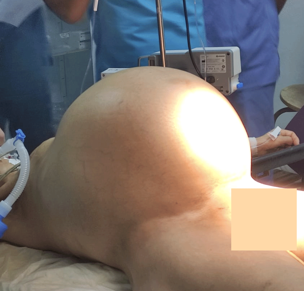
She was 156 cm tall and weighed 65 kg. Despite her apparent body weight, the patient appeared cachectic, with marked cutaneous and mucosal pallor. In striking contrast, abdominal examination revealed massive distension, with an abdominal circumference of 120 cm. The abdomen was tense and protruded above the level of the chest in the supine position. Prominent dilated superficial abdominal veins were visible. Bilateral lower-limb edema was also noted.
The remainder of the physical examination was unremarkable.
Laboratory investigations revealed moderate anemia with a hemoglobin level of 9.6 g/dL. Renal function tests showed urea and creatinine values within the lower limits of normal. Coagulation studies demonstrated a reduced prothrombin time (PT) of 61%. Total serum protein level was elevated at 86 g/L.
Serum tumor markers, including CA-125, CA 19–9, CA 15–3, alpha-fetoprotein (AFP), and carcinoembryonic antigen (CEA), were all within normal reference ranges.
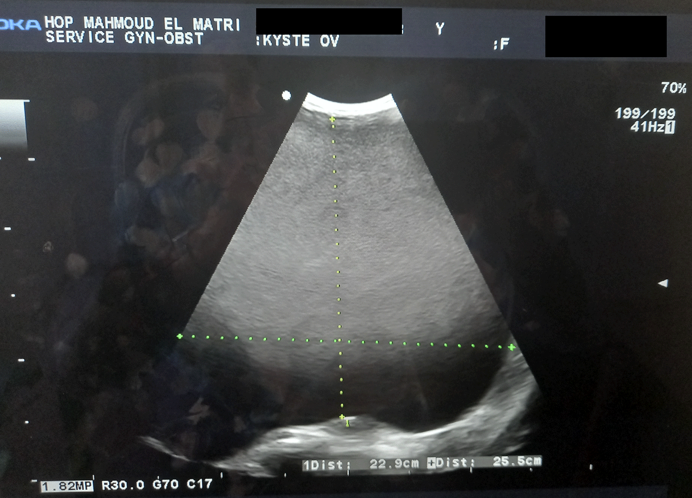
Abdominal and pelvic ultrasonography was limited by the extreme size of the lesion, which could not be entirely visualized within the ultrasound field of view (Figure 1).
Magnetic resonance imaging (MRI) revealed a giant unilocular cystic abdominopelvic mass measuring 37 × 28 × 24 cm, most likely arising from the right ovary. The left ovary was described as follicular in appearance and without abnormal findings (Figures 2, 3).
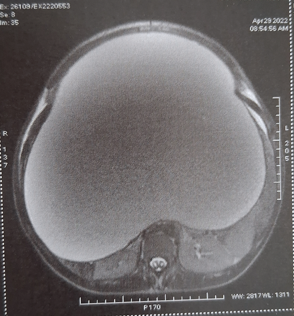
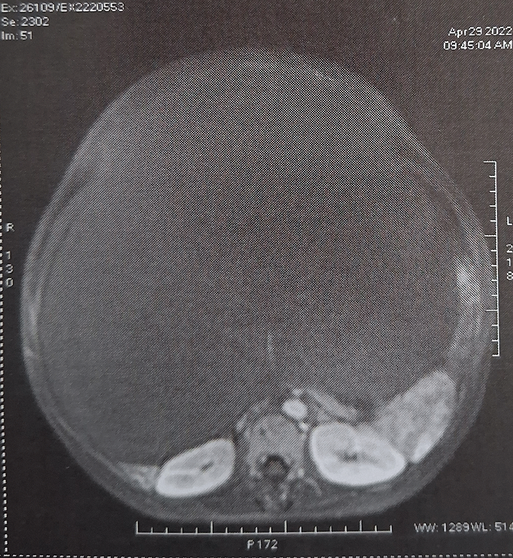
Overall, the imaging and biological findings were suggestive of a benign ovarian cystic tumor.
The patient underwent a midline laparotomy through a subumbilical incision to access the abdominal cavity. Upon entry, a giant cystic mass was encountered (Figures 4, 5).
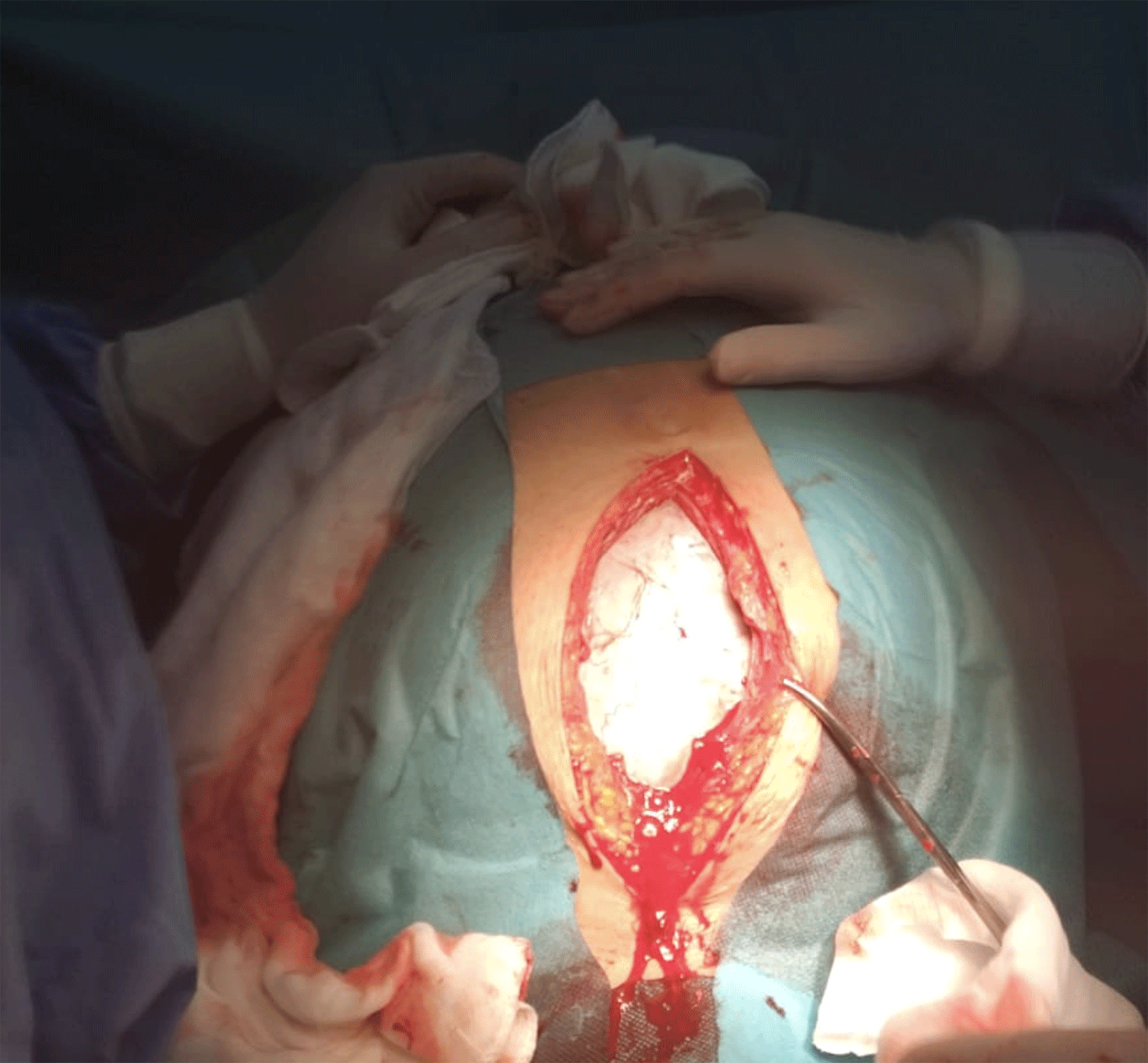
Peritoneal cytology was first performed. Subsequently, the tumor wall was carefully incised, allowing aspiration of approximately 21 liters of thick greenish fluid.
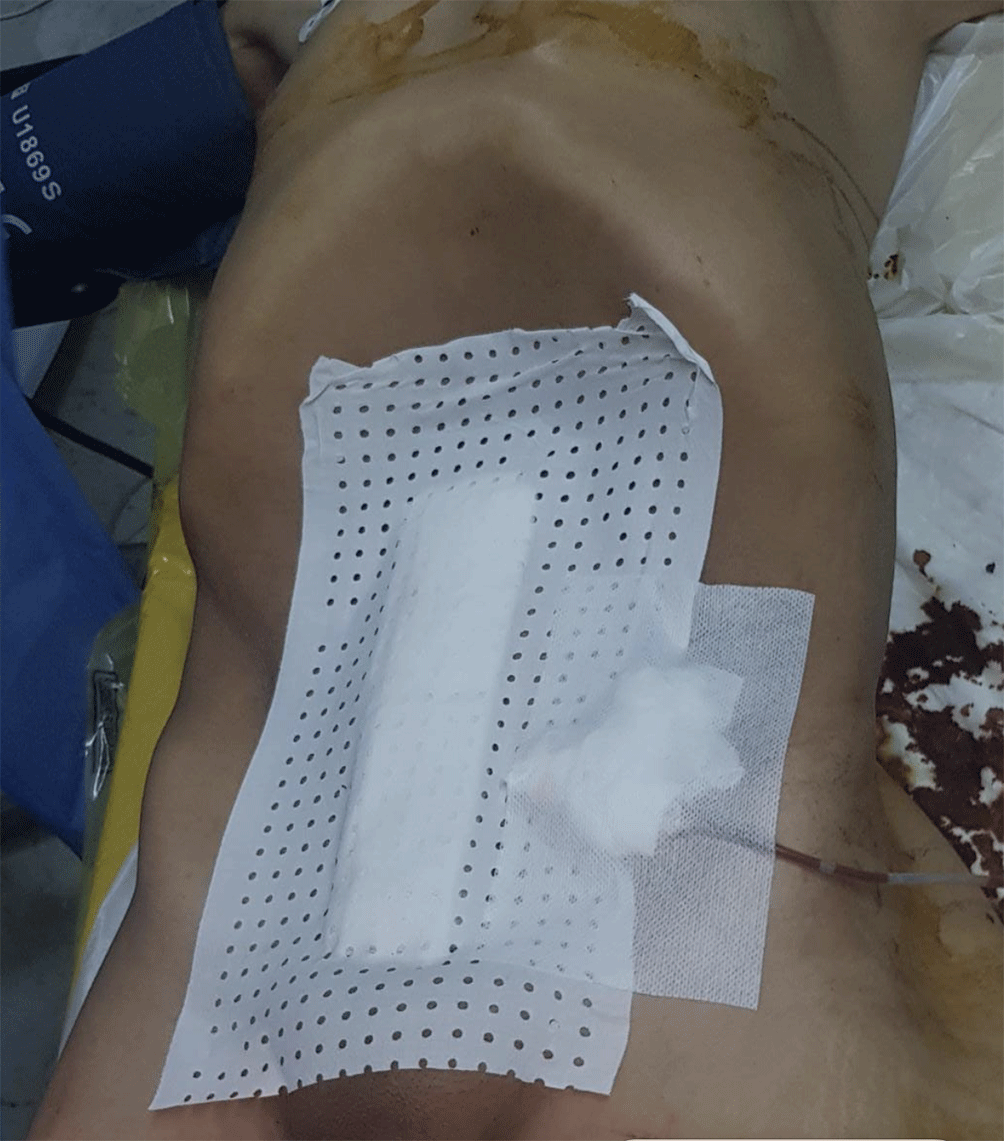
Following decompression of the mass, a systematic exploration of the abdominal cavity was performed. Intraoperative findings confirmed a right ovarian origin of the tumor. The right infundibulopelvic (lombo-ovarian) ligament and utero-ovarian ligament appeared thickened and elongated. Careful inspection of the abdominal and pelvic organs revealed no associated abnormalities. The left ovary, left fallopian tube, uterus, and the remaining abdominal structures appeared grossly normal (Figures 6, 7).
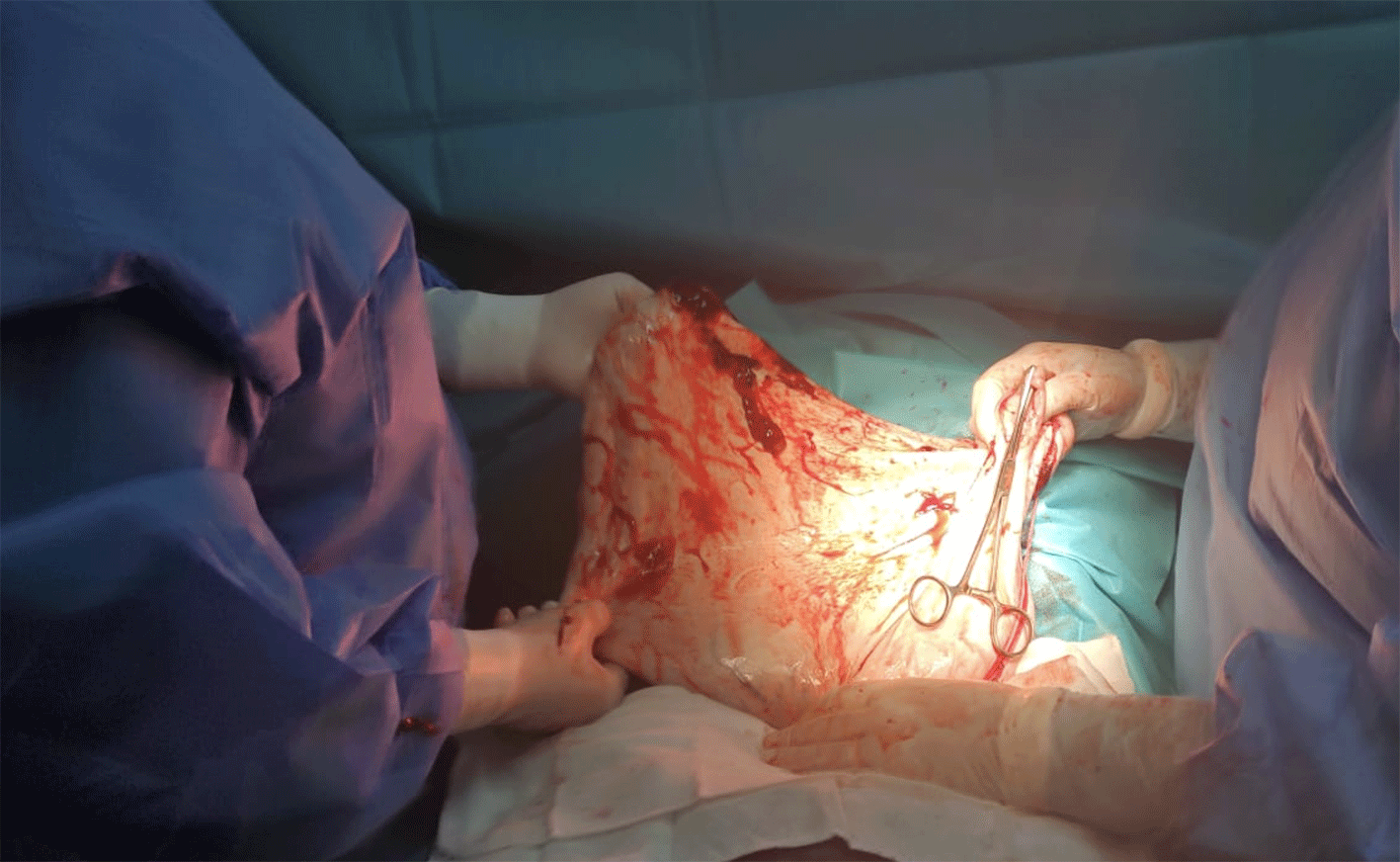
A right adnexectomy was then performed.
Intraoperatively, the patient received transfusion of 2 units of compatible packed red blood cells and 3 units of fresh frozen plasma. The procedure was well tolerated.
In the perioperative period, the patient received intravenous antibiotic therapy and thromboprophylaxis with enoxaparin 40 mg.
Postoperatively, the patient developed hypoalbuminemia with a serum albumin level of 25 g/L, which required correction with appropriate medical management.
The postoperative course was uneventful, with progressive clinical improvement. Recovery was marked by normalization of oral intake, gradual weight gain, and complete resolution of functional complaints. The patient regained normal daily activities without difficulty.
Biological follow-up demonstrated correction of postoperative hypoalbuminemia under supportive medical management.
The patient was discharged in good general condition after an uncomplicated hospital stay.
Histopathological examination of the surgical specimen revealed a borderline mucinous ovarian tumor of endocervical-type, with no evidence of invasive malignancy.
At follow-up, the patient remained asymptomatic, with no clinical or radiological signs of recurrence.
the timeline is detailed in Table 1.
Giant abdominopelvic masses have become increasingly rare in countries with widespread access to gynecological care and diagnostic imaging. Nevertheless, when they occur, they continue to pose significant diagnostic and therapeutic challenges because of their size, the compression of adjacent structures, and the potential for intraoperative complications.5–7
In our patient, the insidious course and exceptional size of the lesion (37 × 28 × 24 cm on MRI) are characteristic of slowly growing ovarian cystic tumors. Such lesions often remain asymptomatic for long periods and may only become clinically apparent once they reach a size sufficient to cause abdominal distension, pelvic discomfort, or gastrointestinal symptoms. The patient’s progressive abdominal enlargement, early satiety, and occasional vomiting were consistent with the mass effect exerted by the tumor.
Serum tumor markers, including CA-125, CA 19–9, CA 15–3, AFP, and CEA, were all within normal reference ranges. Although normal tumor marker levels may support a benign etiology, they do not exclude malignancy, particularly in borderline ovarian tumors.8,9 CA-125, one of the most commonly used biomarkers in the evaluation of ovarian neoplasms, may also be elevated in several benign gynecological conditions, such as endometriosis.8 Therefore, tumor marker assessment should always be interpreted in conjunction with clinical and radiological findings.
Imaging played a crucial role in the diagnostic workup. Although abdominal and pelvic ultrasonography was limited by the enormous size of the lesion, it provided an initial assessment. MRI subsequently enabled a more comprehensive evaluation, demonstrating a giant unilocular cystic mass without solid components, papillary projections, or multiple septations, features generally suggestive of a benign ovarian lesion.10
MRI criteria for differentiating benign from malignant ovarian masses are well established. A unilocular lesion with homogeneous fluid content, a thin and smooth wall, and the absence of mural nodules or vegetations are highly predictive of benign pathology.11 In the present case, these imaging findings initially favored a benign diagnosis.
The decision to perform a laparotomy rather than a laparoscopic approach was dictated by the exceptional size of the mass. Indeed, adnexal masses larger than 15 cm are generally managed through an open approach, allowing complete abdominal exploration, safer hemostatic control, and specimen removal without fragmentation, thereby minimizing the risk of tumor dissemination in cases of unexpected malignancy.12
The surgical procedure consisted of a midline laparotomy, controlled decompression of the cystic mass while preventing intraperitoneal spillage, followed by thorough exploration of the abdominal cavity and right adnexectomy. This strategy allowed safe removal of the mass while preserving optimal surgical conditions and minimizing the risk of intraoperative complications.
This case presents several noteworthy features. First, the extraordinary size of the tumor is rarely encountered in modern practice. Second, beyond its local compressive effects, the mass was associated with a marked deterioration in the patient’s general condition, including significant weight loss, impaired mobility, and nutritional compromise. Such systemic consequences are uncommon but may occur in giant ovarian tumors because of prolonged compression, reduced oral intake, and increased metabolic demands. This observation underscores the potential impact of delayed presentation on both quality of life and overall health status.13
Finally, the absence of suspicious intraoperative findings, the unilocular appearance of the lesion, and normal tumor marker levels initially suggested a benign ovarian cystic tumor. However, histopathological examination ultimately revealed a borderline mucinous tumor of endocervical type, underscoring the importance of definitive pathological assessment for accurate diagnosis and management.
Written informed consent for publication of the patient’s clinical information and accompanying images was obtained from the patient. Copies of the signed informed consent forms in both Arabic and French have been submitted to the journal as supporting documentation. All reasonable efforts have been made to protect the patient’s anonymity; however, complete anonymity cannot be guaranteed.
ChatGPT, OpenAI was used solely as a language and writing assistance tool to improve the clarity, grammar, style, and organization of the manuscript. The AI tool was not used to generate, analyze, interpret, or modify any clinical data, results, images, or scientific conclusions. All clinical information, data collection, analysis, interpretation, and final manuscript content were reviewed and validated by the authors, who take full responsibility for the accuracy and integrity of the work.
The authors would like to thank all healthcare professionals involved in the patient’s care and management.
The author(s) declared that no grants were involved in supporting this work.
© 2026 Mejri O et al. This is an open access article distributed under the terms of the Creative Commons Attribution License, which permits unrestricted use, distribution, and reproduction in any medium, provided the original work is properly cited.
Current Reviewer Status: ?
Key to Reviewer Statuses VIEW HIDE
ApprovedThe paper is scientifically sound in its current form and only minor, if any, improvements are suggested
Approved with reservations A number of small changes, sometimes more significant revisions are required to address specific details and improve the papers academic merit.
Not approvedFundamental flaws in the paper seriously undermine the findings and conclusions
Version 1
VERSION 1
PUBLISHED 31 Jul 2026
Reviewer Report 05 Aug 2026
Marko Bašković, Catholic University of Croatia, Zagreb, Croatia
Not Approved
VIEWS 0
Is the background of the case’s history and progression described in sufficient detail?
Partly
Are enough details provided of any physical examination and diagnostic tests, treatment given and outcomes?
Partly
Is sufficient discussion included of the importance of the findings and their relevance to future understanding of disease processes, diagnosis or treatment?
No
Is the case presented with sufficient detail to be useful for other practitioners?
Partly
Competing Interests: No competing interests were disclosed.
Reviewer Expertise: surgery
CloseOpen Peer Review
Alongside their report, reviewers assign a status to the article:
| Invited Reviewers | |
|---|---|
| 1 | |
| Version 1 31 Jul 26 | read |
Sign up for content alerts
Alongside their report, reviewers assign a status to the article:
Approved - the paper is scientifically sound in its current form and only minor, if any, improvements are suggested
Approved with reservations - A number of small changes, sometimes more significant revisions are required to address specific details and improve the papers academic merit.
Not approved - fundamental flaws in the paper seriously undermine the findings and conclusions