Просмотр новости
Найдите то, что Вас интересует
 Спутники
 РН
РН SpaceX
 Роскосмос
 NASA
NASA
СпутникиРНSpaceXРоскосмосNASABackground Helminth therapy holds considerable theoretical promise for clinical and public health applications. Unfortunately, clinical trials have often used helminths produced using methods that are both highly variable and insufficiently described in the literature, which may hinder reproducibility and troubleshooting. This study aimed to devise a method for purification of helminths that avoids mechanical filtration, which may damage some organisms. In particular, the goal was to devise a method to separate large quantities of cysticercoids of the cestode species Hymenolepis diminuta (HDCs), a process that has not been described previously but that would be necessary before HDCs can be used clinically. Methods The physical size and behavior of HDCs in solution were assessed and used to create a patented washing device. Results The cross-sectional area of HDCs varied over a 10-fold range, from approximately 0.06 to 0.6 mm2, dependent in large part upon the host. That cross-sectional area correlated in a linear fashion with the steady-state speed of HDCs while falling through solution, which varied over a 5-fold range, from approximately 0.4 mm/s to 2.0 mm/s in the solution tested. These parameters were used to develop a washing device that utilized a variable flow speed dictated by changing column diameter and an upward flowing column of liquid that effectively and simultaneously suspended HDCs of various sizes in a wash solution. Conclusions This study describes a detailed method of purification of HDCs, organisms that could potentially be used for helminth therapy. It is hoped that government agencies or non-profit organizations can use such technology to conduct rigorous clinical trials, potentially leading to the production of reasonably priced therapeutics.

Venkatakrishnan A, Swanson C, Palkar A et al. Isolation of Hymenolepis diminuta cysticercoids for therapeutic use: The need for open-source methodology [version 1; peer review: awaiting peer review]. F1000Research 2026, 15:1334 (https://doi.org/10.12688/f1000research.186809.1)
Research Article
[version 1; peer review: awaiting peer review]
Aarushi Venkatakrishnan1, Chelsea Swanson1,2, Antara Palkar1, [...] David Takamiya1, Joshua T. Sarafian1, Jennifer Mauney2, Margaret W. Gatongi3, Jasmine Cendejas Hernandez1, Victoria Lawton1, Zoie E. Holzknecht
https://orcid.org/0009-0000-6012-162X
1,2, John P. Jones III2, Zacharoula Konsoula2, Kateřina Jirků4,5, Garin Aglietti2, Detlev Goj6, William Parkerhttps://orcid.org/0000-0003-3644-9152
1,2,7Aarushi Venkatakrishnan1, Chelsea Swanson1,2, [...] Antara Palkar1, David Takamiya1, Joshua T. Sarafian1, Jennifer Mauney2, Margaret W. Gatongi3, Jasmine Cendejas Hernandez1, Victoria Lawton1, Zoie E. Holzknecht
https://orcid.org/0009-0000-6012-162X
1,2, John P. Jones III2, Zacharoula Konsoula2, Kateřina Jirků4,5, Garin Aglietti2, Detlev Goj6, William Parkerhttps://orcid.org/0000-0003-3644-9152
1,2,71 Surgery, Duke University Medical Center, Durham, NC, 27710, USA
2 WPLab, Durham, NC, 27705, USA
3 Duke Pratt School of Engineering, Durham, NC, USA
4 Institute of Parasitology, Czech Academy of Sciences, Branišovská, Czech Republic
5 University of South Bohemia, Branišovská, Czech Republic
6 Tanawisa Company Ltd, Pranakornsriayuthaya, Thailand
7 Psychology and Neuroscience, The University of North Carolina at Chapel Hill, Chapel Hill, North Carolina, USA
Aarushi Venkatakrishnan
Roles: Data Curation, Formal Analysis, Investigation, Visualization, Writing – Review & Editing
Chelsea Swanson
Roles: Data Curation, Formal Analysis, Investigation, Project Administration, Validation, Visualization, Writing – Original Draft Preparation, Writing – Review & Editing
Antara Palkar
Roles: Data Curation, Investigation, Visualization, Writing – Review & Editing
David Takamiya
Roles: Data Curation, Formal Analysis, Investigation, Writing – Review & Editing
Joshua T. Sarafian
Roles: Data Curation, Investigation, Visualization, Writing – Review & Editing
Jennifer Mauney
Roles: Data Curation, Formal Analysis, Investigation, Methodology, Validation, Writing – Review & Editing
Margaret W. Gatongi
Roles: Conceptualization, Investigation, Methodology, Project Administration, Resources, Software, Supervision, Writing – Review & Editing
Jasmine Cendejas Hernandez
Roles: Investigation, Supervision, Visualization, Writing – Review & Editing
Victoria Lawton
Roles: Data Curation, Formal Analysis, Investigation, Visualization, Writing – Review & Editing
Zoie E. Holzknecht
Roles: Conceptualization, Project Administration, Supervision, Visualization, Writing – Review & Editing
John P. Jones III
Roles: Conceptualization, Formal Analysis, Investigation, Methodology, Validation, Writing – Review & Editing
Zacharoula Konsoula
Roles: Conceptualization, Formal Analysis, Methodology, Validation, Visualization, Writing – Review & Editing
Kateřina Jirků
Roles: Conceptualization, Methodology, Validation, Visualization, Writing – Original Draft Preparation, Writing – Review & Editing
Garin Aglietti
Roles: Conceptualization, Investigation, Writing – Original Draft Preparation, Writing – Review & Editing
Detlev Goj
Roles: Conceptualization, Formal Analysis, Investigation, Resources, Validation, Visualization, Writing – Review & Editing
William Parker
Roles: Conceptualization, Data Curation, Formal Analysis, Funding Acquisition, Investigation, Methodology, Project Administration, Resources, Supervision, Visualization, Writing – Original Draft Preparation, Writing – Review & Editing
OPEN PEER REVIEW
REVIEWER STATUS AWAITING PEER REVIEW
Helminth therapy holds considerable theoretical promise for clinical and public health applications. Unfortunately, clinical trials have often used helminths produced using methods that are both highly variable and insufficiently described in the literature, which may hinder reproducibility and troubleshooting. This study aimed to devise a method for purification of helminths that avoids mechanical filtration, which may damage some organisms. In particular, the goal was to devise a method to separate large quantities of cysticercoids of the cestode species Hymenolepis diminuta (HDCs), a process that has not been described previously but that would be necessary before HDCs can be used clinically.
MethodsThe physical size and behavior of HDCs in solution were assessed and used to create a patented washing device.
ResultsThe cross-sectional area of HDCs varied over a 10-fold range, from approximately 0.06 to 0.6 mm2, dependent in large part upon the host. That cross-sectional area correlated in a linear fashion with the steady-state speed of HDCs while falling through solution, which varied over a 5-fold range, from approximately 0.4 mm/s to 2.0 mm/s in the solution tested. These parameters were used to develop a washing device that utilized a variable flow speed dictated by changing column diameter and an upward flowing column of liquid that effectively and simultaneously suspended HDCs of various sizes in a wash solution.
ConclusionsThis study describes a detailed method of purification of HDCs, organisms that could potentially be used for helminth therapy. It is hoped that government agencies or non-profit organizations can use such technology to conduct rigorous clinical trials, potentially leading to the production of reasonably priced therapeutics.
allergic, autoimmune, cestode, helminth therapy, purification
Corresponding author: William Parker Competing interests: Coauthors MWG, JCH, and CS are currently employees of Duke University. Duke University owns intellectual property described in this manuscript and could benefit financially if therapeutic use of HDCs gains regulatory approval. In addition, coauthor MMG has financial interests in the intellectual property described in this manuscript. Finally, all authors have experience/expertise with either regulatory issues associated with the clinical application of helminth therapy, and/or the production of helminths under controlled conditions, and thus have advantages for employment and financial gain should helminth therapy gain regulatory approval.
Grant information: This work was funded by the employers of the authors, particularly Duke University Medical Center, Tanawisa, WPLab and the Czech Academy of Sciences. Coauthors GA and WP are CEOs of Tanawisa (for profit) and WPLab (nonprofit), respectively.
Copyright: © 2026 Venkatakrishnan A et al. This is an open access article distributed under the terms of the Creative Commons Attribution License, which permits unrestricted use, distribution, and reproduction in any medium, provided the original work is properly cited. How to cite: Venkatakrishnan A, Swanson C, Palkar A et al. Isolation of Hymenolepis diminuta cysticercoids for therapeutic use: The need for open-source methodology [version 1; peer review: awaiting peer review]. F1000Research 2026, 15:1334 (https://doi.org/10.12688/f1000research.186809.1) First published: 08 Aug 2026, 15:1334 (https://doi.org/10.12688/f1000research.186809.1) Latest published: 08 Aug 2026, 15:1334 (https://doi.org/10.12688/f1000research.186809.1)
The concept of “biota restoration” involves the reintroduction of organisms into the ecosystem of the human body which have been lost as a result of “systems hygiene”, or changes imposed by human civilization aimed at preventing infectious disease.1 These changes have resulted in the loss of contact with a variety of symbiotic organisms, primarily various types of helminths, including roundworms (nematodes) and tapeworms (cestodes). Health problems associated with this loss, termed biota depletion, include allergic, autoimmune and neuropsychiatric conditions.2,3 Helminth therapy, therefore, serves as a first step in biota restoration by treating individuals with immunological problems associated with biota depletion. In addition, some helminths may attenuate the development of colon cancer under some conditions.4–7 However, progress in the field of helminth therapy has lagged, potentially due in part to issues associated with inconsistencies in production of therapeutic organisms.8 Such inconsistencies are particularly evident in trials utilizing ova from the porcine whipworm, Trichuris suis (TSO).
Most clinical trials in the field of helminth therapy have been conducted with TSO, with mixed results. Some studies have shown success,9–13 while others failed14–17 or yielded equivocal results.18–20 Unfortunately, detailed methods describing production of TSO for clinical trials are absent from the literature, leaving questions regarding whether differences in production protocols may have affected results.8,21 As an example of the paucity of methodological details available, out of 12 trials with TSO that have been reported,9–20 none described the food supplied to the porcine hosts, and only four described any details regarding the porcine hosts. Three studies described “pathogen-free pigs”,10–12 without further detail, and another study described “barrier-bred Gottingen minipigs (miniature pigs)”.17 Only two studies clearly describe viability of the ova used in the trials,9,13 and only one study described how the TSO were administered to the porcine hosts for production.10
The information that has been published regarding production of TSO for clinical trials indicates that production methods have varied dramatically over time. For example, some trials employed ova produced in vitro, 10–12 whereas others employed ova produced in vivo.9,17 At least two different washing methods for in vitro production have been described, one involving a potassium dichromate solution,12 and the other involving a phosphate buffered solution.10,11 Storage of TSO for clinical trials has also been variable, with solutions described as saline,12 phosphate buffered saline,10,11 sulfate-stabilized 0.05 mol/L H2SO4 (pH 1.0),17 phosphoric acid buffer, pH 5, with 0.05% potassium sorbate preservative,9,18 with storage up to 2.5 years at 2-8 °C,9 a solution at pH 5 with preservative,14 or simply a “liquid”,15,16 depending on the study.
Clinical trials have been conducted with two nematodes, including TSO,9–20 a whipworm which imbeds itself into the gut epithelium, and larva of the hookworm Necator americanus, 22–28 which penetrate the skin, burrow through the lungs, and then penetrate the gut epithelium of the small bowel. However, no trials have yet been reported with a cestode or with a species of helminth that remains within the lumen of the gut. Nevertheless, socio-medical studies evaluating the therapeutic potential of the cestode Hymenolepis diminuta, an organism that remains within the lumen of the gut, have shown that this organism may be effective, particularly for treatment of some allergic and neuropsychiatric conditions.21,29
We have previously described a detailed production method for Hymenolepis diminuta cysticercoids (HDCs),30 the immature stage of Hymenolepis diminuta that can be cultivated in a laboratory and potentially used for helminth therapy. For such cultivation, adult H. diminuta are maintained in laboratory rats (Rattus norvegicus), and the cysticercoids can be maintained in a variety of insects, although grain beetles (Tenebrio molitor) are most often used in the laboratory. The method we previously described30 was based upon a consensus among producers of the organisms for use in self-treatment-based helminth therapy. Although the HDCs live in a sterile environment in the extraintestinal space of their beetle hosts, bacterial contamination occurs when the HDCs are extracted from the host. Although bacteria present in grain beetles, an edible insect, are not hazardous to humans, growth of those microorganisms in storage solutions could potentially damage HDCs and lead to a rancid/unpalatable product. Further, bacterial growth would likely prevent the product from meeting FDA requirements for bacterial burdens in non-sterile pharmaceutical preparations.31 Bacteria are readily removed from the HDCs by washing, but handling of individual HDCs during the washing process is time consuming to the point of prohibiting utilization of the organisms in sufficient quantities for clinical trials.30
HDCs are morphologically complex, containing a juvenile helminth in their core, surrounded by a flexible, relatively fragile jelly-like layer and a tail-like structure that is not part of the juvenile helminth, but rather part of the jelly-like layer that is shed when the juvenile helminths excyst (emerge from the cysticercoid).32,33 The HDCs’ jelly-like outer coating, variable size, and flat, disk shaped geometry present unique challenges when attempting to separate the organisms from bacterial contaminants. Here, we present a novel, scalable protocol for washing HDCs that preserves their morphology and viability. This includes the use of a patented device specifically designed to accommodate the organisms’ unique geometry and structural fragility.
The overall goal of the study was to develop a method of washing bacterial contaminants from HDCs on a large scale. A washing device utilizing liquid flow and gravity to create a steady state washing condition (see the section entitled “Washing device”, below) was envisioned to wash the HDCs en masse. Prior observations made in our laboratory30 indicated that substantial differences in the size of HDCs exist, and it was hypothesized that these size differences may affect the organisms’ terminal velocities while falling through a solution, which would in turn affect the design parameters necessary for the washing device. With that in mind, the first phase of the study involved an assessment of (a) size variation in HDCs and (b) the corresponding speed (rate) at which the HDCs fall through solution. The second phase of the study involved the design, production and use of a device to wash multiple HDCs at a given time. The design and operation of the device was guided in large part by variations in HDC size and fall rate determined in the initial phase of the study. Since the intent of the work was not to evaluate biological variation in HDC size within the population, but rather to develop a method of isolating all HDCs within a population, hosts (grain beetles) or HDCs were selected non-randomly to ensure that a mixture of relatively small and large organisms were evaluated. This approach facilitated determination of the relationships between HDC size and fall rate. However, to determine the association between individual hosts and HDC size, HDCs were necessarily selected for evaluation at random from a given host. Random selection was carried out by selecting the first organisms observed under the microscope. During selection, organisms which had a damaged (broken, not intact) tail-like structure were excluded and not evaluated.
For measurements of fall rates as a function of HDC size, HDCs were selected so that a broad distribution of sizes were included in the sample. Measurements were halted at n = 28 after it was determined that a sufficient sample set had been obtained to draw conclusions regarding the connection between sample size and fall rate. For assessment of HCD size within a given beetle, 10 to 13 HDCs were selected at random from each beetle for detailed measurement. Beetles were selected so that a broad distribution of HDC sizes was obtained, and experiments were halted when it was determined that a sufficient sample set had been obtained to draw conclusions regarding the association between beetle host and HDC size. For assessment of washing device function, beetles were selected at random and sufficient HDCs were extracted to load the device with between 80 and 120 HDCs for each run. The position of individual HDCs were assessed in the wash chamber in order to evaluate device function, and thus the number of HDCs Loaded in the device at a given time was limited by the ability to identify individual HDCs in the wash chamber: overcrowding in the chamber made it difficult to identify individual HDCs in the chamber.
Assessment of HDC viability after washing were conducted using 20 to 40 organisms, which was a convenient sample size for assessment in a single day. Since 100% of the HDCs were viable after washing, it was determined that experiments using HDCs without washing were unnecessary. No data points were excluded from the study.
The use of small sample sizes (10 to 13 organisms) for measurements of HDC size (2 dimensional surface area) as a function of the beetle host was selected as a starting point based on qualitative observations that HDC size tended to vary between hosts more so than within a given host. Since these observations were borne out while conducting the study (see Results), the use of larger samples sizes proved unnecessary. Correlations between continuous variables (size and fall speed) were evaluated using linear regression, and comparisons of size between different hosts were evaluated using ANOVA. GraphPad Prism 5 (by Dotmatics) was used for statistical analyses. A p-value < 0.05 was considered significant.
The use of laboratory rats as primary hosts for Hymenolepis diminuta was approved by the Duke University Animal Care and Use Committee (protocols A022-16-02 and A275-18-12). The Duke University Institutional Review Board (IRB) approved the application for patent coverage for the technology described in this manuscript (United States US Patent Application No. 17/296,908) by coauthor WP and, at the same time, the conduct of related research on human subjects by coauthor WP under the condition that co-author WP irrevocably refuse any potential financial benefits from the patent. That research on human subjects conducted by coauthor WP and colleagues (Duke IRB protocol Pro00045035) has been completed and published,8 and is not the subject of the original work described in this manuscript.
HDCs were cultured as previously described using Sprague Dawley rats as primary hosts and Tenebrio molitor (grain beetles) as secondary hosts.30 Under these conditions, laboratory rats host H. diminuta for the duration of their natural life span without apparent adverse effects from the tapeworms, and are sacrificed only when a humane endpoint is reached due to a general age-related decline in health or other age-associated issues (e.g. breast tumor development) unrelated to the experimental protocol. Clarified almond milk, produced as previously described,30 was used for all procedures involving handling of isolated HDCs, unless otherwise indicated. Imaging software (Leica Application Suite V4.5; Leica Microsystems, Switzerland) and a Zeiss Stemi 2000-C microscope with a Leica MC 120 HD camera and a Zeiss KL 1500 LCD light source were used to photograph HDCs, and, ImageJ image analysis software (ImageJ 1.51k, Wayne Rasband, National Institutes of Health, USA) was used to quantify the size of individual HDCs. Heads and tails of HDCs were quantified separately (see results). Fall speed through solution was measured over a 20 cm distance in a 6 mm ID glass column using a stopwatch.
A glass washing device was fabricated (Carolina Research Glass Inc., Greensboro, NC) as shown in Figure 1 in order to wash HDCs of various sizes without contact with filters. The primary innovation is the “wash-hold” section (258 mm in length) with a diameter that increases from bottom (6 mm ID) to top (15 mm ID), as shown in the figure. As wash fluid is pumped from the inlet port up to the outlet port, appropriate adjustment of flow rates (see Results) results in the suspension of HDCs in the wash-hold section. HDCs can be eluted from the bottom of the device or, alternatively, a harvesting probe can be inserted into the wash-hold section ( Figure 1). For this study, all washed HDCs were harvested from the bottom of the device. The device has been patented (US patent 12,409,193 B2, September 9th, 2025).34
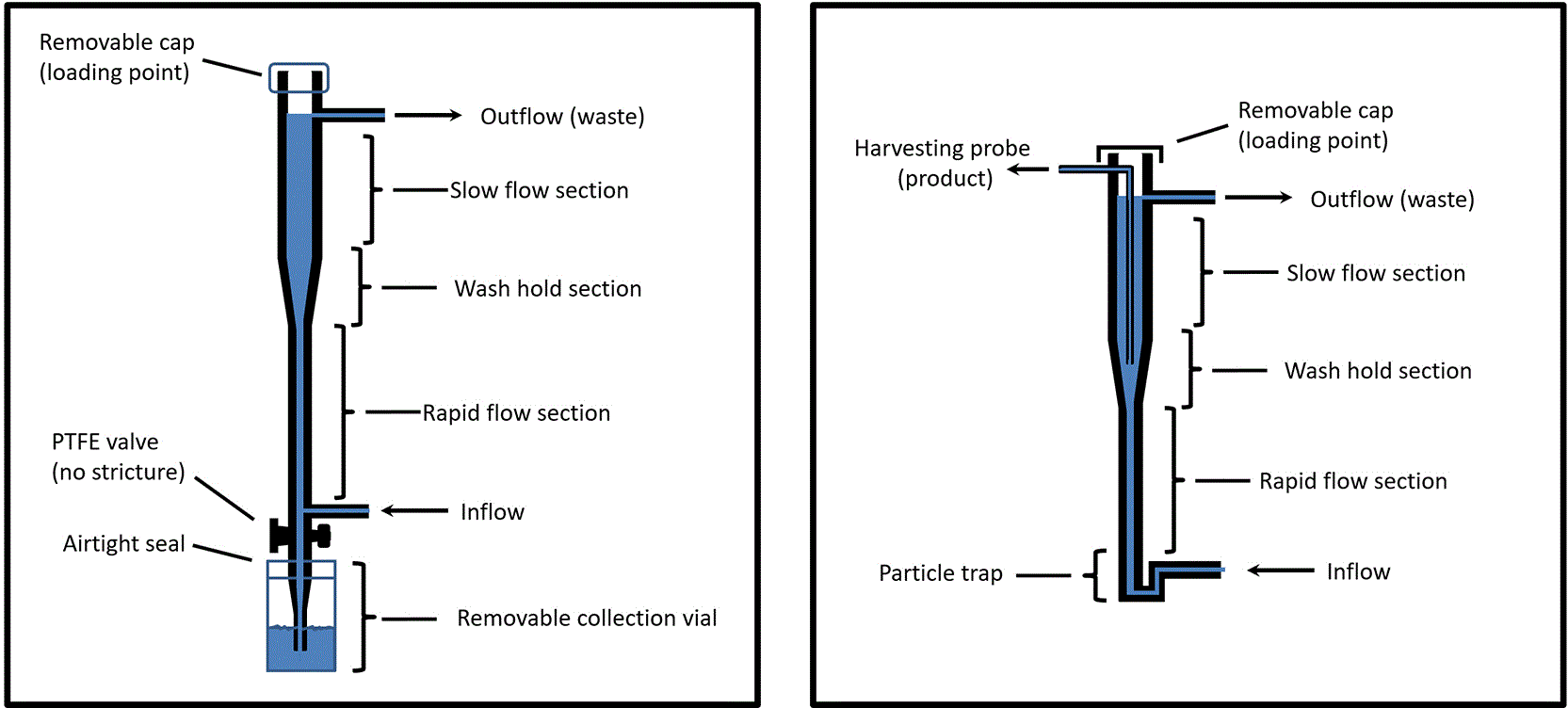
HDC’s can be harvested by decreasing the flow speed to > 0.04 cm/s in the rapid flow section, which enables elution of the HDCs from the bottom of the device on the left. On the right, insertion of a harvesting probe from the top into the wash-hold section is shown as an option. The harvesting probe allows for collection of any fraction of the HDCs in the wash-hold section rather than 100% of the HDCs in the wash-hold section, and allows the insertion of a particle trap in the event that any large contaminants are present in the HDC preparation.
Viability of the HDCs was determined based on an excysting assay performed using an ibidi Stage Top Incubator (ibidi GmbH, Grafelfing, Germany) at 37 °C. For the assay, HDCs were placed in a 96 well plate (lumox® multiwell, 96, Sarstedtstr, D-51588 Nümbrecht Germany) using a Samco 231 disposable fine-tip pipette. Clarified almond milk was removed using the pipette, and 100 μl of 5 mg/ml pepsin, 60 mM HCl and 0.425% saline in 50% Tyrode’s saline buffer warmed to 37 °C was added to each well. The plate was then covered with an ibidi glass cover and observed at 37 °C for 10 minutes. The cover was then removed and the HDC’s were washed with approximately 300 μl of unbuffered 0.54% saline. The saline solution was removed, and 150 μl of 5 mg/ml trypsin with 5 mg/ml tauroglycocholate in 20 ml of Tyrode’s saline warmed to 37 °C was added to each well. Excysting was monitored for 90 minutes.
The size of HDCs was found to vary over a considerable range based on approximations of the cross-sectional area of the organisms when lying flat on a dish in solution ( Figure 2). Head size was well correlated with tail size ( Figure 2), with the total estimated cross-sectional area (head size + tail size) varying over a 10-fold range ( Figure 3). The estimated area in the cross section was strongly dependent on the particular grain beetle (Tenebrio molitor, the secondary host) from which the HDCs were extracted ( Figure 3) for reasons that are unknown.
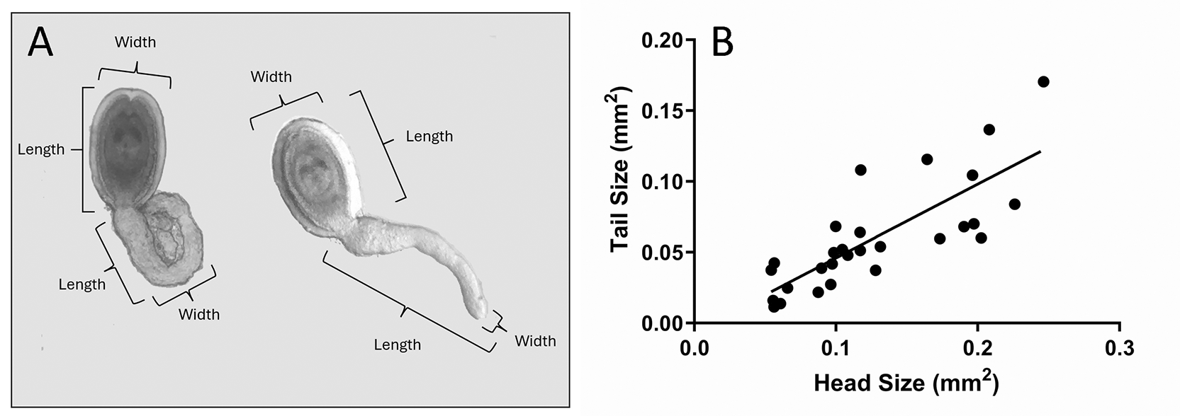
(A) The cross sectional area of each HDC was approximated by multiplying the length by the width for head and tails independently, then adding the areas together. Two HDCs, one with a compact (“ball shaped”) tail and one with a long tail, are shown for illustration. In the photo, background debris in the micrograph has been removed and the photos adjusted in size and contrast to match for the illustration. (B) Comparison between the two measurements, head size and tail size showed a positive correlation (C, n=28, r2= 0.6236, p<0.0001). The least-squares regression line is shown.
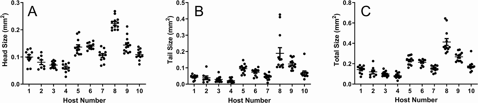
Variable size of HDCs as a function of the grain beetle (Tenebrio molitor, the secondary host) from which the HDCs were extracted. The head size (A), tail size (B), and total size (C) were assessed. n1= 10, n2= 8, n3= 12, n4= 10, n5= 12, n6= 10, n7= 11, n8= 12, n9= 13, n10= 11. With a one-way Anova test, p<0.0001 for A, B, and C. The bars indicate means and standard errors.
The fall speed of HDCs in solution varied over approximately a 5-fold range, and was dependent on helminth size as assessed by estimated cross-sectional area when lying flat on a dish in solution ( Figure 4). In addition to the effect of total cross-sectional area on fall speed, the effect of the shape of the tail on the fall speed was evaluated. The tail shape of HDCs is highly variable, with some having a long, extended tail, whereas others contain a more compact tail ( Figure 2, left panel).30 Based on comparison of the fall speed of HDCs with straight tails to that of HDCs with more compact tails, the shape of the tail did not apparently affect the fall speed ( Figure 4D).
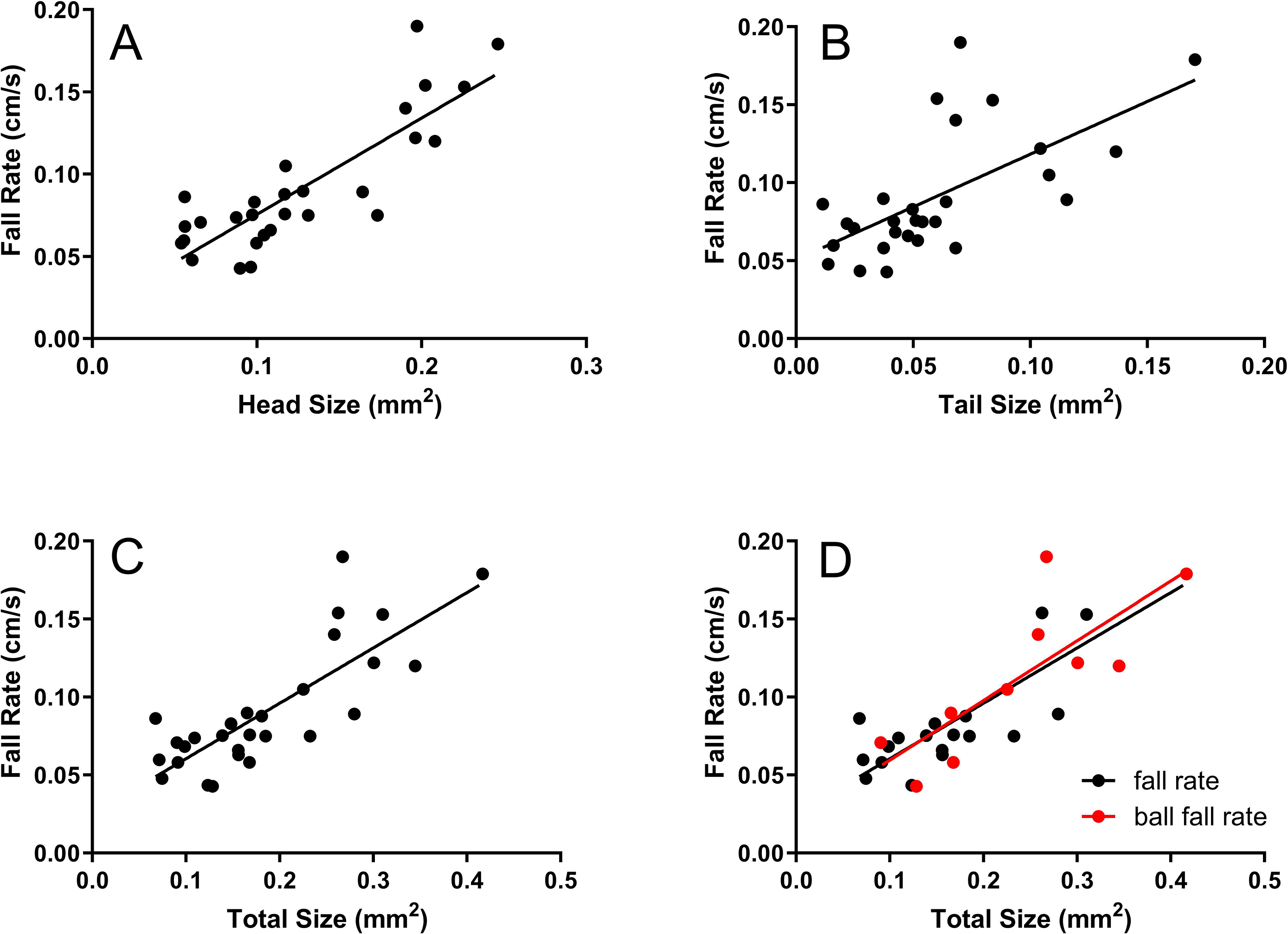
The least-squares regression lines are shown.
The washing device described in the Methods ( Figure 1) effectively held HDCs in a steady position as the organisms “fell” through solution ( Figure 5). The location of the organisms in the wash-hold section was linearly related to the flow rate ( Figure 5). After 90 minutes of washing, viability was assessed via excystation as described in the Methods. 100% of HDCs successfully excysted immediately after washing (n=31). Furthermore, 100% (n=23) remained viable and excysted after being held in saline for an additional 24 hours post wash, demonstrating viability of the organisms following the washing procedure.
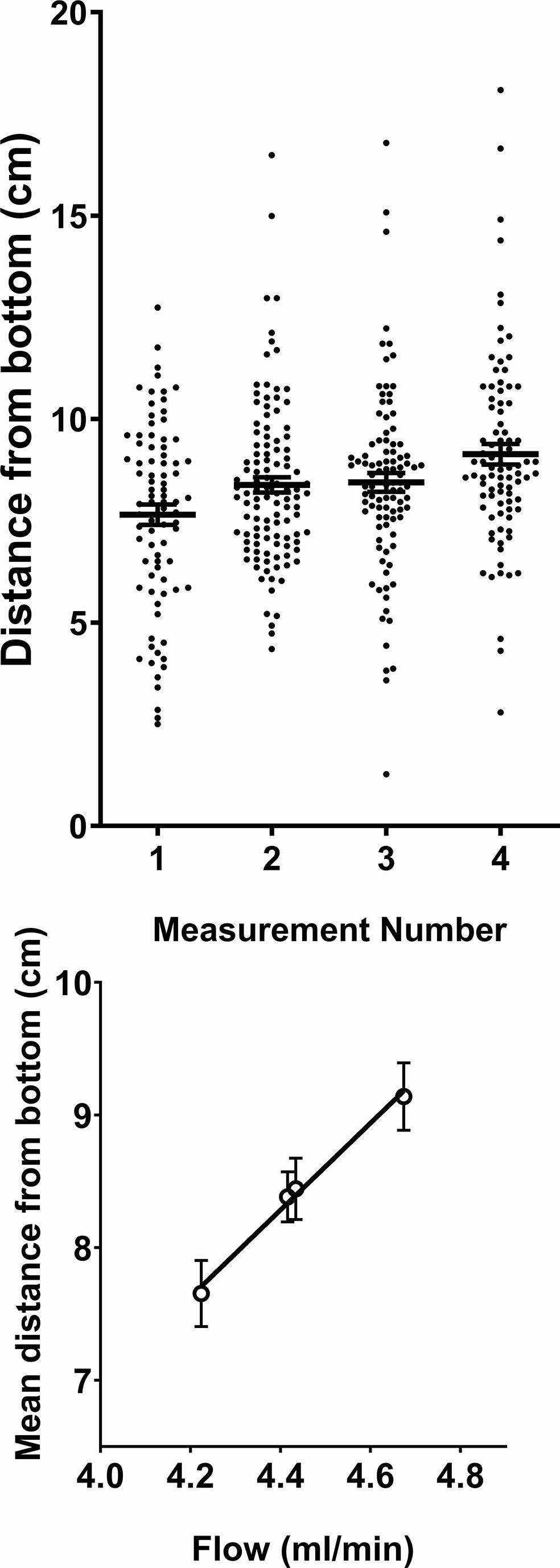
The distance was measured from the bottom of the wash-hold section. Four measurements were taken with three different flow rates (measurements 2 and 3 used the same flow rate). The number of organisms assessed in measurements one through four was 84, 111, 97, and 88, respectively. The top panel shows measures for individual HDCs, and the bottom panel shows average measures for each run as a function of flow rate. The mean distance from the bottom of the wash-hold section correlated strongly with the flow rate in a linear fashion (n=4, r2= 0.9918, p=0.0041). The least-squares regression line is shown, and the means and standard errors are indicated.
This paper presents a “no contact” washing procedure for HDCs, designed to effectively overcome limitations in the production of HDCs for therapeutic use that were previously described by our group.30 This approach is sufficient to prepare large numbers of HDCs for clinical use in a manner that meets current FDA guidelines for bacterial contamination. Notably, HDCs remain viable after this washing procedure based on their ability to excyst. However, the viability of washed HDCs under long-term storage conditions remains an important area for future work. Additionally, it remains unknown whether an assessment of excysting accurately reflects the therapeutic potential of HDCs in humans, and whether some therapeutic potential might be lost during washing or storage regardless of whether the organisms can excyst. These questions are particularly relevant for industrial-scale production and application. In the absence of validated biomarkers to assess the therapeutic function of HDCs in vitro, the only way to gain answers to the questions above will likely be through clinical trials.
Despite all these limitations, Biome reconstitution remains a critical step towards alleviating underlying causes of chronic inflammatory diseases and normalizing immune function in Western society.2,3 However, as noted in the Introduction, utilizing a product in clinical trials for which the production method is highly variable and not publicly available imposes considerable limitations. First and foremost, without access to a production protocol, reproducibility by others is impossible. Second, if results are inconsistent, tracing the source of variability can be difficult. Troubleshooting has been particularly difficult in the field of helminth therapy, as companies have folded due to failure of clinical trials and academic investigators have retired without publishing detailed protocols. These challenges underscore the need for an open-source method for the production of helminths used in clinical trials. However, this criterion presents challenges for commercial development, as patenting a species of helminth for clinical applications may prove difficult,35–37 leaving producers to rely on trade secrets for protection of their investments.
The quandary faced by investigators hoping to develop helminth therapy for widespread clinical use arises from a conflict between the considerable theoretical potential for helminths to normalize immune function and the practical challenge of protecting commercial interests necessary to procure funding of resource-intensive clinical trials from the commercial sector. One feasible solution is for governmental organizations and/or well-funded non-profits to conduct the clinical trials using an open-source approach. FDA approval of a product not protected by exclusive patent rights is conceivably an approach that could open the field for commercial development and yield dramatic benefits for both clinical practice and public health.
This study was not preregistered with a data analysis plan at an independent registry. The ARRIVE checklist is available at Parker, William (2026), “Helminth purification data v1”, Mendeley Data, V2, doi:10.17632/dmht2t9tdr.238
Data, graphs, and results of statistical tests are available at Parker, William (2026), “Helminth purification data v1”, Mendeley Data, V3, doi:10.17632/dmht2t9tdr.338
Coauthor WP is grateful to the Duke Center for Translation and Commercialization, to the Duke IRB, and to the Duke IACUC for assistance and advice, without which this work would not have been possible.
Coauthors MWG, JCH, and CS are currently employees of Duke University. Duke University owns intellectual property described in this manuscript and could benefit financially if therapeutic use of HDCs gains regulatory approval. In addition, coauthor MMG has financial interests in the intellectual property described in this manuscript. Finally, all authors have experience/expertise with either regulatory issues associated with the clinical application of helminth therapy, and/or the production of helminths under controlled conditions, and thus have advantages for employment and financial gain should helminth therapy gain regulatory approval.
This work was funded by the employers of the authors, particularly Duke University Medical Center, Tanawisa, WPLab and the Czech Academy of Sciences. Coauthors GA and WP are CEOs of Tanawisa (for profit) and WPLab (nonprofit), respectively.
© 2026 Venkatakrishnan A et al. This is an open access article distributed under the terms of the Creative Commons Attribution License, which permits unrestricted use, distribution, and reproduction in any medium, provided the original work is properly cited.
Current Reviewer Status:
AWAITING PEER REVIEW
AWAITING PEER REVIEW
?
Key to Reviewer Statuses VIEW HIDE
ApprovedThe paper is scientifically sound in its current form and only minor, if any, improvements are suggested
Approved with reservations A number of small changes, sometimes more significant revisions are required to address specific details and improve the papers academic merit.
Not approvedFundamental flaws in the paper seriously undermine the findings and conclusions